Type of image used for interproximal examination – Interproximal examination plays a crucial role in dentistry, and the type of image used can significantly impact the accuracy and effectiveness of the examination. This article delves into the various types of images employed for interproximal examination, exploring their advantages, limitations, and diagnostic value.
From bitewings to panoramic radiographs, each imaging technique offers unique insights into the interproximal region. Understanding the strengths and weaknesses of these imaging modalities empowers dental professionals to select the most appropriate approach for each patient, ensuring optimal diagnosis and treatment outcomes.
Interproximal Examination Image Types: Type Of Image Used For Interproximal Examination

Interproximal examination is a crucial aspect of dental diagnosis, as it allows dentists to assess the health of the teeth and their surrounding structures. Various types of images are used for this purpose, each with its own advantages and limitations.
Bitewing Images
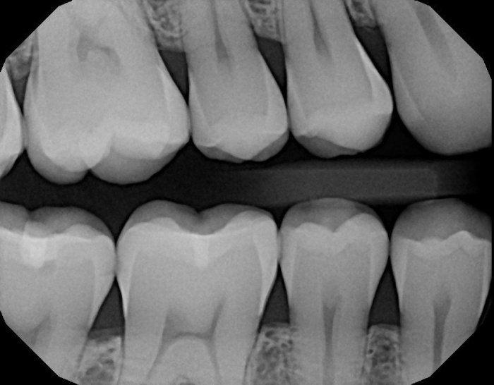
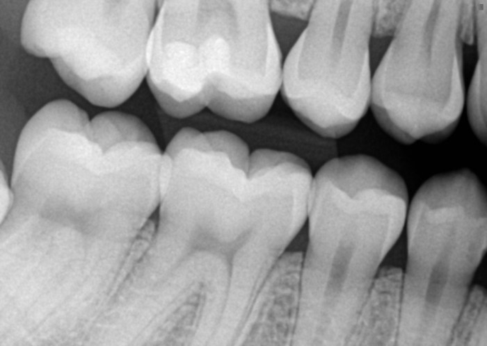
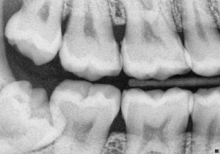
Bitewing images are one of the most commonly used types of images for interproximal examination. They are taken with a small x-ray machine placed inside the patient’s mouth. Bitewings provide a clear view of the crowns and roots of the teeth, as well as the interproximal spaces between them.
This makes them ideal for detecting caries, periodontal disease, and other interproximal pathologies.
Periapical Images
Periapical images are another type of image used for interproximal examination. They are taken with a larger x-ray machine placed outside the patient’s mouth. Periapical images provide a detailed view of the entire tooth, including the roots and the surrounding bone.
This makes them useful for diagnosing apical lesions, root fractures, and other periapical pathologies.
Panoramic Radiographs
Panoramic radiographs are a type of dental x-ray that provides a wide-angle view of the entire mouth. They are taken with a machine that rotates around the patient’s head. Panoramic radiographs are useful for screening for interproximal caries and other dental problems, but they are not as detailed as bitewing or periapical images.
Digital vs. Traditional Images, Type of image used for interproximal examination
Digital images are becoming increasingly popular for interproximal examination. They offer several advantages over traditional film-based images, including improved image quality, reduced radiation exposure, and the ability to be stored and shared electronically. However, digital images can be more expensive than traditional images, and they require specialized equipment.
Advanced Imaging Techniques
In some cases, advanced imaging techniques such as cone beam computed tomography (CBCT) and magnetic resonance imaging (MRI) may be used for interproximal examination. These techniques provide even more detailed images than traditional x-rays, which can be useful for diagnosing complex dental problems.
Commonly Asked Questions
What is the most common type of image used for interproximal examination?
Bitewing images are the most commonly used type of image for interproximal examination.
What are the advantages of using panoramic radiographs for interproximal examination?
Panoramic radiographs provide a wide field of view, making them useful for screening for interproximal caries and other pathologies.
What are the limitations of using periapical images for interproximal examination?
Periapical images have limited diagnostic value for interproximal caries detection due to their narrow field of view.

